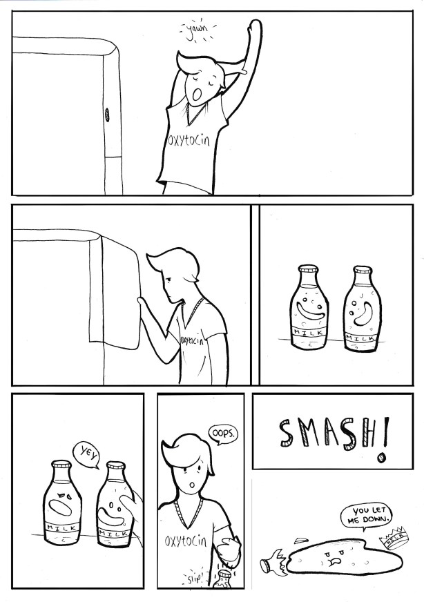
A reminder from sensational artist Sarah Mills, that whilst it is Prolactin which promotes milk production in the female, it is Oxytocin which lets milk down…
A reminder from sensational artist Sarah Mills, that whilst it is Prolactin which promotes milk production in the female, it is Oxytocin which lets milk down…
There are six basic nutrient groups. Three of them provide energy and three are non-energy nutrients.
Energy Nutrients
Non-Energy Nutrients
There are three forms of carbohydrates:
Sugars – Provide energy quickly, they are relatively simple molecules and are absorbed and used efficiently by the body – glucose is the most important simple sugar.
Starches – Provide energy more slowly as they are longer chains which must first be broken down into simple sugars before they can be used.

Fibre – Fibre is an indigestible form of carbohydrate in cats and dogs. Rabbits, Horses and Ruminants (like cattle and sheep) can break down fibre to use as energy. Cats and dogs cannot break down fibre and so it can be used to bulk out the diet and control an animal’s weight by making the animal feel full without providing lots of energy.
A common quandry in parasitology is identification of different types of parasites.
When it comes to the subject of lice, there are some happy coincidences which make life easier.
For Sucking Lice, simply remember the initial letter ‘S‘ :
:
They Suck
They have Small heads
They move Slowly
They have Sideways or forward pointing antennae
They are around 5mm in size
(ok, stretching it but the number ‘5’ looks a wee bit like an ‘S’)
For Biting Lice, the letter ‘B‘ is all important:

They Bite
They have Big heads
They Move Bloomin’ quick (ahem!)
They have Backward pointing antennae
They are around 3mm in size
(if you draw a vertical line through the start of the number ‘3’, it becomes a ‘B’)
The body contains many tubes for carrying substances from A to B, for example

Peristaltic Contractions in the gut. (Wikipedia)
Rarely does the body rely on gravity in order to move substances through these tubes, instead it relies on the contractions of the muscle in the wall of the organ – a process called peristalsis.
All of these tubes mentioned contain a special kind of muscle in their walls, called Smooth Muscle (this is different from the skeletal or striated muscle attached to the skeleton and different to the cardiac muscle which is found in the heart).
When food, for example, enters the intestine from the stomach, the muscle at the start of the intestine will contract, squeezing the bolus of food a bit further along the intestine.
As the food bolus is squeezed along, the next bit of intestine will then contract, moving the food bolus even further. At the same time, the first bit of intestine stops squeezing, so it looks like there is a wave of muscle contractions moving along the length of the intestine.
This process is found all over the body where there are hollow ‘tubes’ responsible for moving substances through the body.
A common cause of confusion in anatomy is the difference between cilia and villi. Both of these are finger-like projections found in the body but they are found in different places and have entirely different jobs to do.
Cilia are tiny hair-like structures which are found on the surface of cells lining the upper-airways (the trachea and bronchi). The job of the cilia is to WIGGLE! The airways are coated with a thin layer of mucus which traps dust and particles stopping them from getting into the lungs. The cilia wiggle to move the mucus up the airways toward the mouth so that it can be coughed up and swallowed.

Villi are finger like structures found in the wall of the intestine [villus = one; villi = many]. Because they come out from the wall of the intestine, they have the effect of creating more space for absorption of nutrients; that is: they increase the surface area. Villi are filled with blood vessels to take away the nutrients to the circulation. They also contain a structure called a lacteal which absorbs fats from the intestine for delivery to the blood stream.

The surface area of the villus is increased even further by the presence of microvilli. Microvilli are tiny structures on the surface of the villi.
NOTE: VILLI DO NOT WIGGLE – THEY DO NOT MOVE FOOD THROUGH THE INTESTINE.
 Remembering the names of the valves of the heart can be difficult – especially trying to remember which valve goes on which side.
You may have heard names like Tricuspid Valve and Bicuspid Valve or even Mitral Valve.
There’s an easier way of naming the valves, however, where the names actually tell you about their position:
Between each Atrium and Ventricle is a valve, which is there to stop blood flowing in the wrong direction. If you use the name Atrio-Ventricular Valve or A-V Valve, the name tells you that the valve is between the Atrium and Ventricle. To make sure everyone knows which side you’re talking about, you should use the names Left Atrio-Ventricular Valve or Right Atrio-Ventricular Valve.
One of the main problems with diarrhoea is that it’s so bloomin’ difficult to spell.
If you have trouble remembering how to spell ‘Diarrhoea’, remember this handy phrase:
Dashing – In – A – Rush? – Really – Hurry – Or – Else – Accident.

The first thing you have to get used to when you see a diagram of the heart (or any other organ) is that they always seem to be ‘back to front’. When you see a picture of the heart, the left is always on the right and the right is always on the left. This is simply because that’s how you would look at someone’s heart if you could look into their chest (or if you had ‘x-ray vision’!). If you’re struggling with this idea, look at your friend’s face and you’ll see that their left ear is on your right and their right ear is on your left.

In mammals, the heart is divided into four chambers with two chambers on the left side and two chambers on the right side. Between the left and right sides of the heart is a wall of muscle called the ‘septum’.
These four chambers have names and that’s the next thing you’ll have to learn.
The same chambers are found on both the left and right side, so you only have two new words to learn which are Atrium and Ventricle.
At the top left of your diagram you’ll see the Right Atrium and underneath it, you’ll see the Right Ventricle. At the top right of your diagram you’ll see the the Left Atrium and under it, the Left Ventricle.
Do remember, it’s only back-to-front on the diagram, the dog’s right ventricle really is on the right side of its heart!
A final tip to help you remember to put the Atrium at the top and the Ventricle at the bottom is to make sure you write the name with a capital letter, draw a line from top to bottom through the first letter of each name and follow the direction of the arrows you’ve just created.

 Cortex and Medulla are words which pop up all over the subject of anatomy. The cortex is always the bit around the outside of a structure and the medulla is always the bit in the middle.
Examples include:
The Renal Cortex – the bit around the outside of the kidney
The Adrenal Cortex – the bit around the outside of the adrenal gland
The Cortex of bones (also known as Cortical Bone) – the thick dense bone tissue around the outside of bones
The Cerebral Cortex – the bit around the outside of the brain
 The Renal Medulla – the bit on the inside of the kidneys
The Adrenal Medulla – the bit on the inside of the adrenal gland
The Medulla of bones (also known as the medullary cavity) – the space in the middle of a long bone where the bone marrow is found
Use the similar sounds of the words MEDULLA and MIDDLE to help you remember!

